Tarah Rhoda: Salt Mine
Artist(s):
Title:
- Salt Mine
Exhibition:
Creation Year:
- 2016
Size:
- 1:50 min.
Category:
Artist Statement:
Nature resonates, resolving forms and echoing solutions similarly at small and large scales. Throughout history man has used these links to contemplate his place in the world. It can even be said that theories of the microcosm and macrocosm emerge from the primitive human instinct of empathy. My work is the result of my personal attempt to explore my connection to the natural world. Investigating the body as an archeological site, literally ‘mining’ myself, I abstract the resulting observations and extractions into poetic reflections and devices, often utilizing laboratory equipment to set the stage.
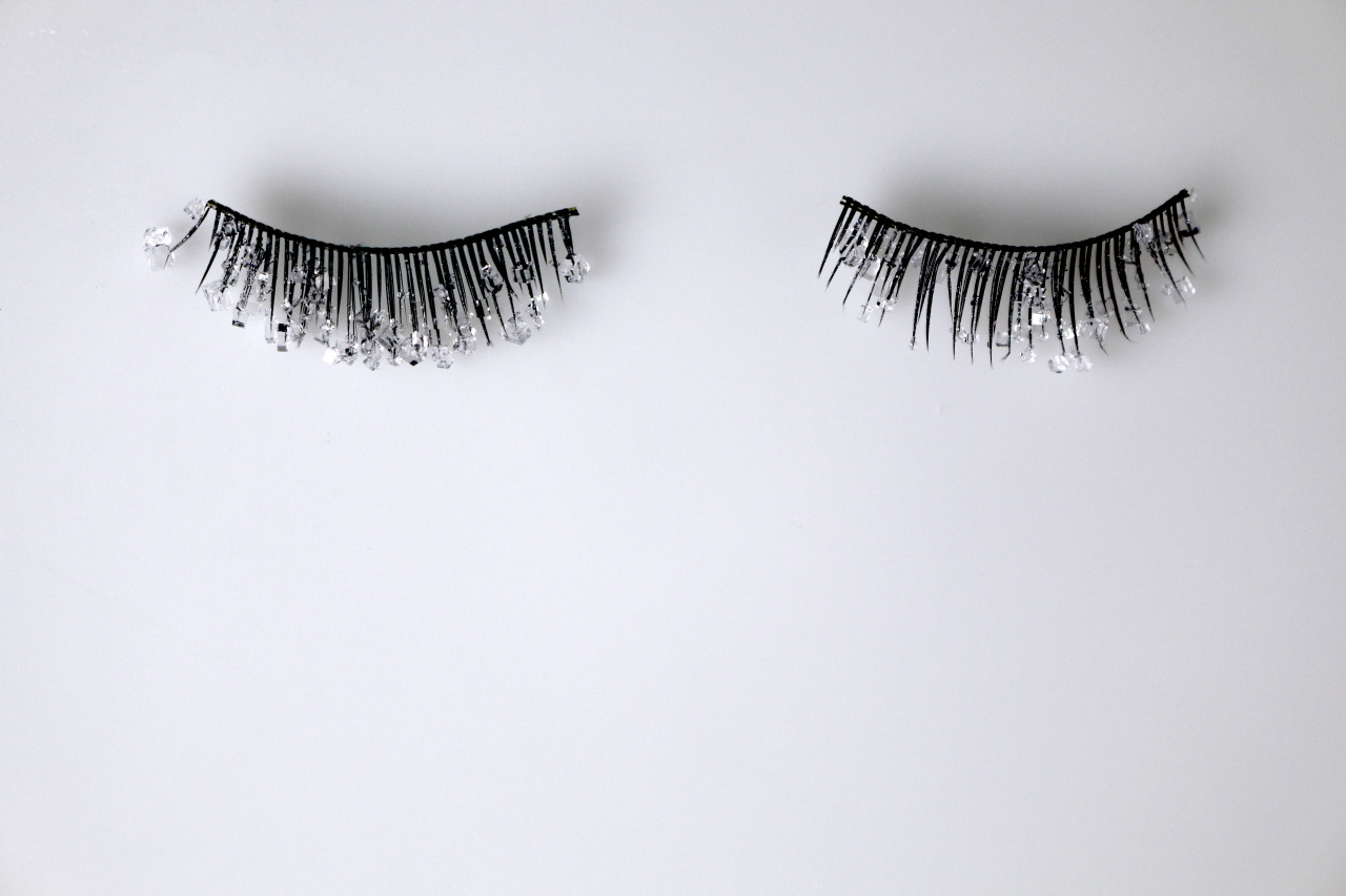
My tear project began in 2012 with the intention of harvesting my own salt by dehydrating tears in petri dishes. I documented all of my samples under compound and inverted microscopes at a range of magnification and was curious to see such variation. I knew the chemical composition of emotional tears had hormones and proteins that weren’t present in basal and reflex tears, but was that what I was seeing? After reviewing over a hundred samples, the differences and similarities were inconclusive. I consulted a lab technician and drafted sterile protocol to control my samples, avoiding cross contamination and other indirect factors. It became clear how the tears were collected had much more to do with the composition of the crystal lattice structure than what “type” of tears they were. Because a solution crystalizes around solid nuclei, debris and particles play a major role in the resulting crystalline structure. If the sample is collected off the cheek, rather than directly from the tear duct it will have excessive skin cells, which will determine the sites of nucleation. If it is the first tear shed, there will be more debris in the duct, rather than if the duct has been flushed.
Technical Information:
Media Used: Compound and inverted microscopes @4X/10X/20X/40X, Canon 5D Mark III, tears.


